The imperative for innovation in histopathology is not merely conceptual—rather, it is rooted in our collective experience. An important case shared by a colleague, involving a missing prostate core from a 12-part biopsy in an otherwise exemplary GU laboratory, illustrates the gravity of the situation. Each core represents a patient’s anticipation of a diagnosis that could alter their life’s trajectory—from benign prostatic hyperplasia (BPH) necessitating lifestyle modifications, to aggressive malignancies with the potential for metastasis and devastating consequences.
The urgency for accurate, timely diagnosis is paramount, and the stakes could not be higher. On this particular day, the pathologist reviewing the slides, noticed a discrepancy: only 11 cores were present. A search ensued, but the missing core could not be found. The histotech was baffled, the pathologist was forced to move on to the mounting caseload, yet the gnawing fear remained: what if that missing core held the key to diagnosis? Some small unspoken dread around “quality of care” lingered back in the mind.
Eventually, the remaining cores were deemed cancer-free. However, this offered false reassurance. Four months later, the patient returned with worsening symptoms. Three months after that, he was gone. A potentially treatable cancer had been missed due to a workflow breakdown.
This devastating outcome, while thankfully rare, is not unheard of in our field. It is a harsh reminder that even the most dedicated professionals are vulnerable to errors within a system that relies heavily on manual processes and fragmented workflows. For pathologists, the weight of such an error is immense, as it directly impacts patient lives and outcomes.
The Imperfect Reality of Traditional Histopathology
This anecdote underscores the broader challenges facing histopathology today. Our workflows, largely untouched by innovation for decades, are riddled with inefficiencies. Consider the multiple handoffs and potential points of failure for a single specimen: from the procedure room requisition to grossing, embedding, sectioning, staining, and finally, to the pathologist’s microscope. Each step introduces the risk of specimen damage, mislabeling, or loss.
Furthermore, studies have shown that nearly half of all errors occur during the critical final stages of reporting, leading to missing or incorrect information in the very document that guides patient care. This reality is echoed in a recent survey revealing a staggering 95% of pathology professionals believe the majority of errors originate within the lab itself.
The TWOD Solution: A Paradigm Shift in Specimen Handling
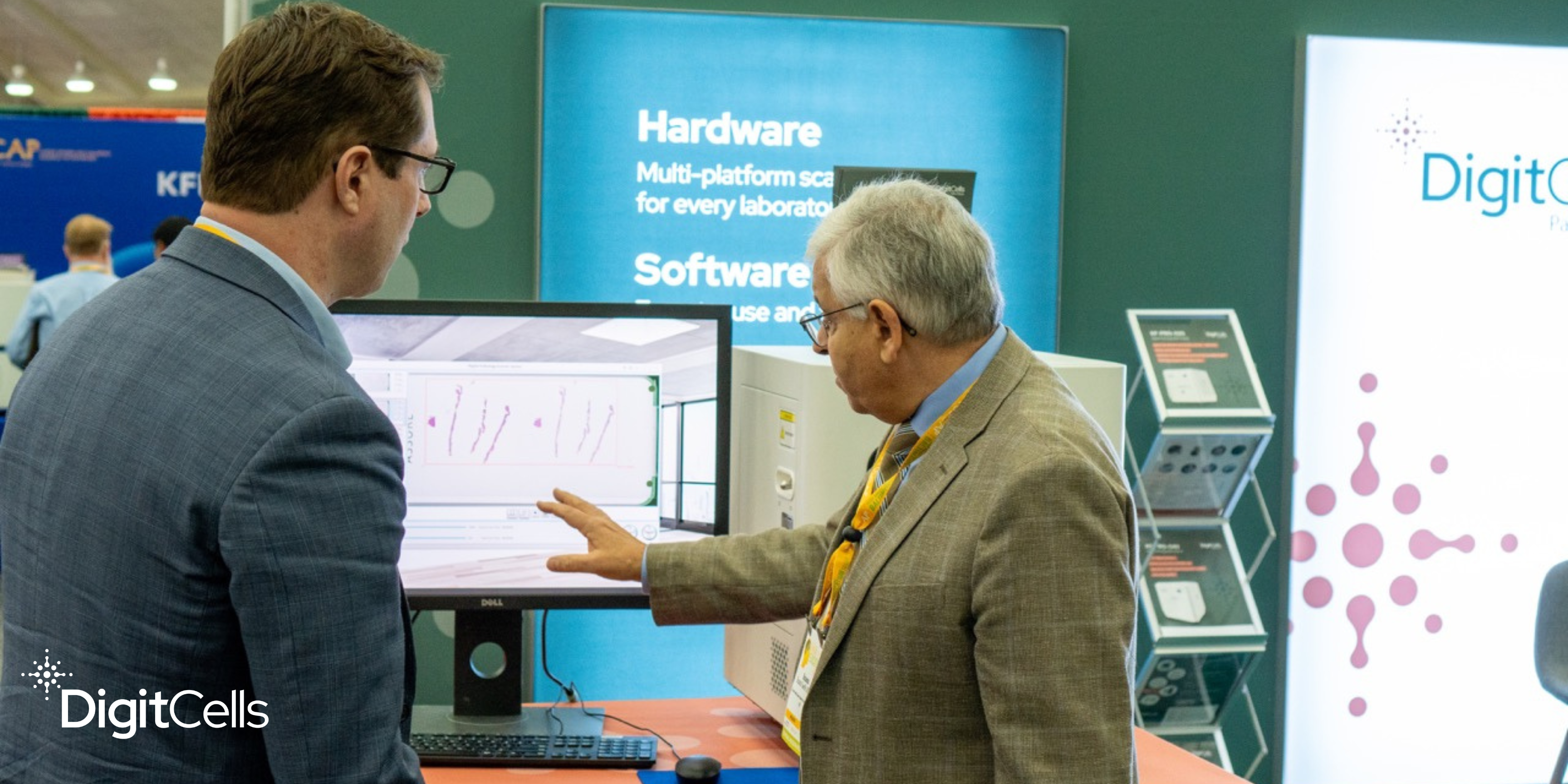
Recognizing the urgent need for change, DigitCells developed the Tissue Workflow Optimization for Digital (TWOD) system. TWOD is not merely a product; it is a comprehensive, end-to-end solution encompassing both a clinician workstation and an AP lab workstation, designed to transform how histopathology is practiced, starting with prostate biopsies.
Key Advantages of TWOD:
- Uncompromised Specimen Integrity from the Source: Our patented cassettes revolutionize specimen handling by enabling clinicians to place biopsy cores directly from the patient into individually barcoded and tracked cassettes. This eliminates the traditional, error-prone transfer process from formalin jars to cassettes, ensuring optimal integrity from the moment of collection.
- End-to-End Traceability: TWOD’s integrated tracking system follows each specimen throughout the entire workflow, from the procedure room to the pathologist’s review. This comprehensive chain of custody minimizes the risk of misidentification or loss, providing clinicians and pathologists with unparalleled confidence in the accuracy and reliability of the procurement and diagnostic process.
- Streamlined Workflow: TWOD consolidates cores, reducing reagent use, staining time, and storage needs. This translates to significant cost savings for labs, while also accelerating turnaround times for faster diagnoses.
- AI-Powered Analysis: TWOD’s rigorously validated algorithms automate and standardize biopsy measurements, ensuring accuracy and objectivity while minimizing manual labor and data entry errors.
- Digitized Slides: TWOD leverages whole slide imaging (WSI) for rapid digital review, enabling pathologists to efficiently assess multiple cores on a single slide. Digital slides also facilitate collaboration and second opinions, further enhancing diagnostic accuracy.
- Seamless Integration: TWOD is designed to seamlessly integrate with existing lab processes, minimizing disruptions and allowing for a smooth transition to digital pathology.
The TWOD Difference: Beyond the Bench
TWOD is not just about technology; it is about empowering clinicians and pathologists together to improve patient care. By addressing the root causes of errors and inefficiencies, TWOD helps to collaborate seamlessly and elevate patient care.
To learn more about how TWOD can revolutionize your lab, contact us at DigitCells. Let’s work together to build a future where histopathology is defined by precision, efficiency, and peace of mind.